
Figure 18.
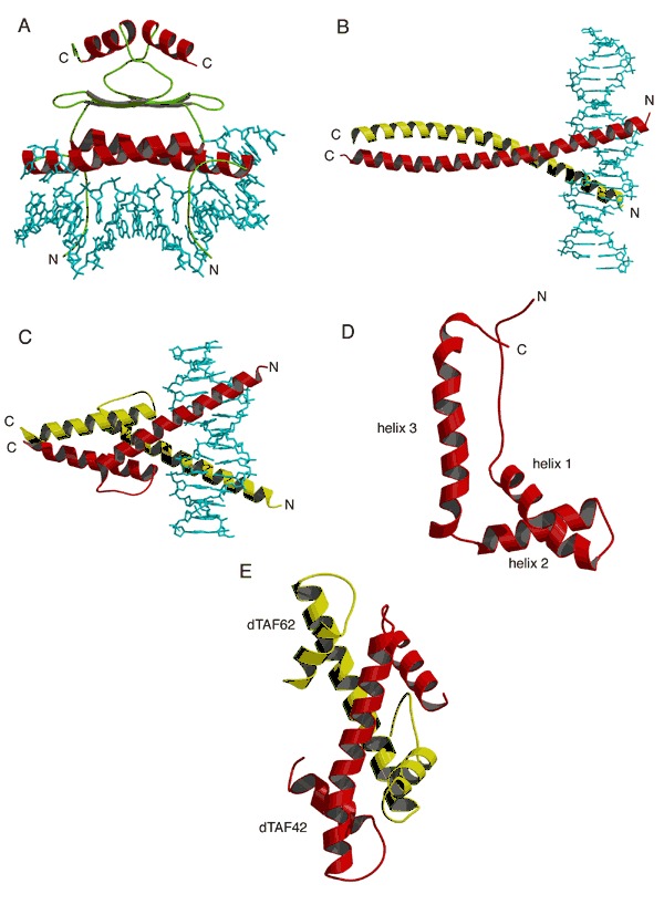
Helical DNA binding domains (A) MADS box of serum response factor bound to DNA (PDB entry 1srs). (B) Basic‐region leucine‐zipper (bZIP) c‐Fos/c‐Jun heterodimer complexed with DNA (PDB entry 1fos). Monomers within the dimer are shaded differently. (C) Basic helix‐loop‐helix (bHLH) MyoD homodimeric transcription activator complexed with DNA (PDB entry 1mdy). Monomers within the dimer are colored differently. (D) High‐mobility group (HMG) fragment B from rat (PDB entry 1hme). (E) Structure of a heterodimer of dTAF42 and dTAF62 (PDB entry 1taf). Each monomer contains the histone fold.