
Figure 19.
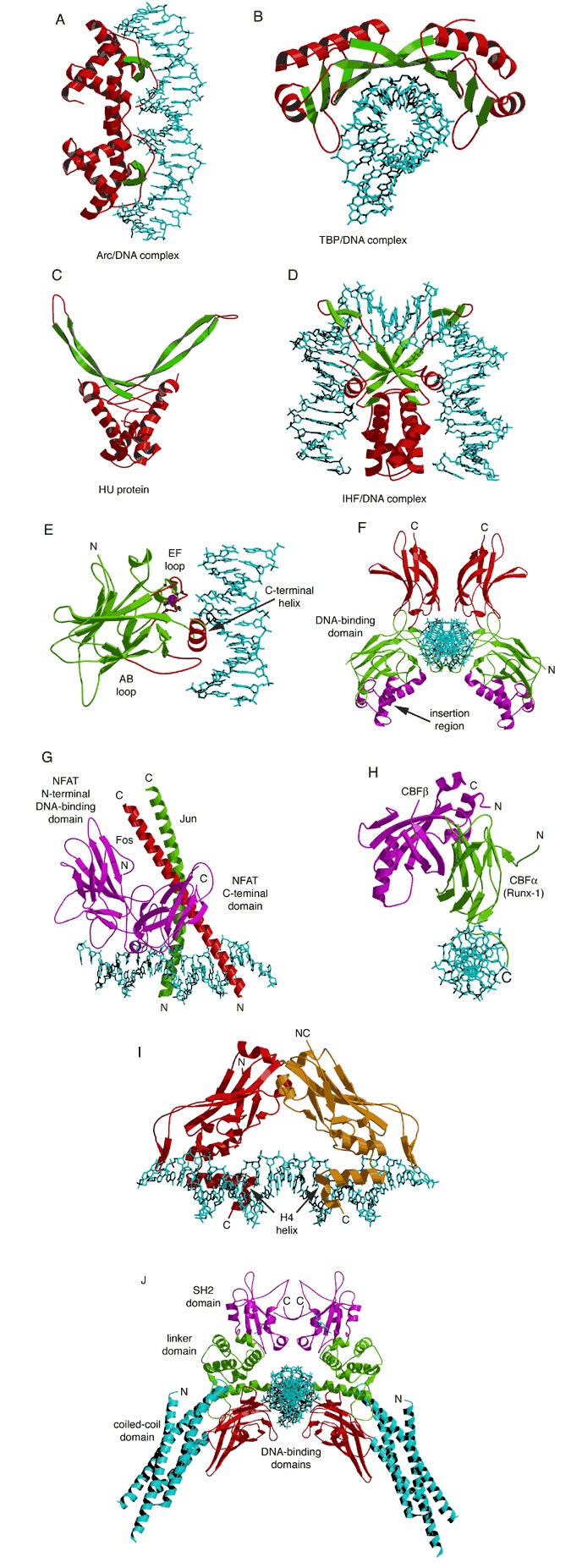
β‐sheet DNA‐binding motifs. (A) Arc‐repressor tetramer complexed with DNA (PDB entry 1par). (B) TATA‐box‐binding protein (TBP) complexed with DNA (PDB entry 1ytb). (C) Histone‐like HU protein (PDB entry 1hue). (D) HU‐like IHF complexed with DNA (PDB entry 1ihf). (E) The p53 tumor‐suppressor monomer bound to DNA (PDB entry 1tsr). Regions involved in DNA binding are labeled and colored red. The zinc atoms and side‐chain ligands to the zinc are represented by ball‐and‐stick models. (F) Structure of the p50/p50 homodimer complexed with DNA (PDB entry 1svc). The two insertion regions (magenta) may play a role in binding other transcription factors. (G) Tetrameric complex of NFAT1 (magenta), Fos/Jun (AP‐1), and DNA (PDB entry 1a02). (H) Ternary complex of CBFα (green), CBFβ (magenta), and DNA (blue) (PDB entry 1h9d). (I) Brachyuri T‐domain homodimer bound to DNA (PDB entry 1h6f). (J) STAT‐1 homodimer bound to DNA (PDB entry 1bf5). The coiled‐coil domain, DNA‐binding domain, linker domain, and SH2 domain are colored blue, red, green, and magenta, respectively.