
Figure 23.
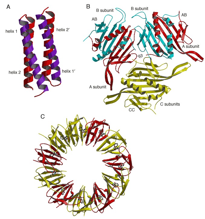
(A) The RNA‐binding protein Rop from E. coli. (PDB entry 1rop). Helices 1 and 1′, postulated to bind to RNA, are labeled. (B) Hexamer of the MS2 phage‐coat protein (PDB entry 1mst). Two AB dimers and a CC dimer are arranged around a quasi‐3‐fold rotation axis located in the center of the figure (labeled q3). (C) Eleven‐subunit oligomer of Trp RNA‐binding attenuation protein (TRAP; PDB entry 1wap). Bound tryptophan molecules are shown by a ball‐and‐stick model. Alternating monomers are shaded differently for clarity.