
Figure 25.
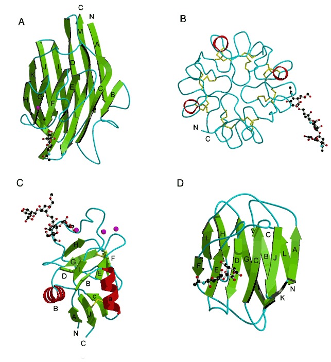
Lectin folds. (A) Legume lectin soybean agglutinin (PDB entry 1sba). The β strands are labeled A through M. The bound carbohydrate molecules are represented by ball‐and‐stick models. (B) Wheat‐germ agglutinin monomer with bound carbohydrate. Disulfide bonds are shown as yellow sticks. (PDB entry 2wgc). (C) The C‐type lectin mannose‐binding protein A (PDB entry 2msb) with bound carbohydrate. Calcium ions are shown as magenta spheres. (D) S‐lectin with bound carbohydrate (PDB entry 1slt).