
Figure 26.
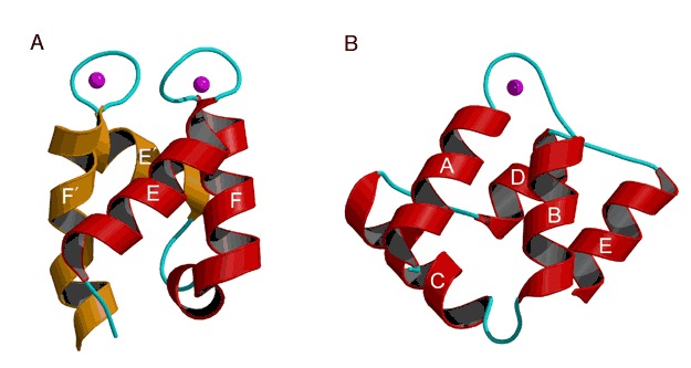
Calcium‐binding folds. Calcium ions are drawn as magenta spheres. (A) A pair of calcium‐binding EF‐hands (red and orange respectively) complexed with Ca2+ from bovine calbindin D9K. The corresponding helices are labeled E, F, E′, and F′ (PDB entry 4icb). (B) Calcium‐binding domain of annexin V (PDB entry 1ala).