
Figure 28.
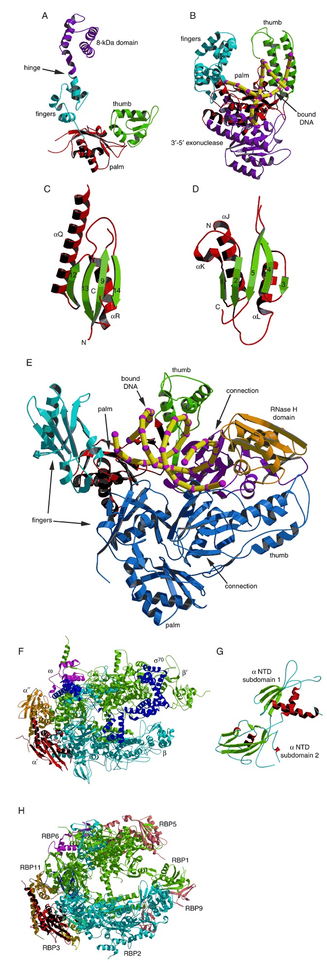
Structures of DNA and RNA polymerases. (A) The 39‐kDa catalytic domain of rat DNA polymerase β (PDB entry: 1bpd). (B) The Klenow fragment of E. coli DNA polymerase I bound to an 11‐bp duplex DNA in an editing complex (PDB entry: 1kln). The DNA is represented by a yellow and purple phosphate backbone trace. (C) The conserved palm subdomain from Klenow fragment (PDB entry 1kln). (D) The palm subdomain from rat DNA polymerase β (PDB entry 1bpd). (E) Structure of the HIV‐1 reverse transcriptase heterodimer complexed with a 19/18‐base duplex DNA (PDB entry 1hmi). The p66 subunit is colored by subdomain and the p51 subunit is dark blue. The bound DNA is represented by a yellow and purple phosphate backbone trace. (F) Thermus thermophilus RNA polymerase holoenzyme (PDB entry 1iw7). The β′, β, α′, α″, ω, and σ70 subunits are labeled and colored green, cyan, red, tan, magenta, and dark blue, respectively. (G) The N‐terminal domain of the RNAP α subunit from E. coli (PDB entry 1bdf). (H) RNAP II from yeast (PDB entry 1i50). Visible subunits are labeled and the five subunits in common with bacterial RNAP follow the same coloring scheme as in (F).