
Figure 32.
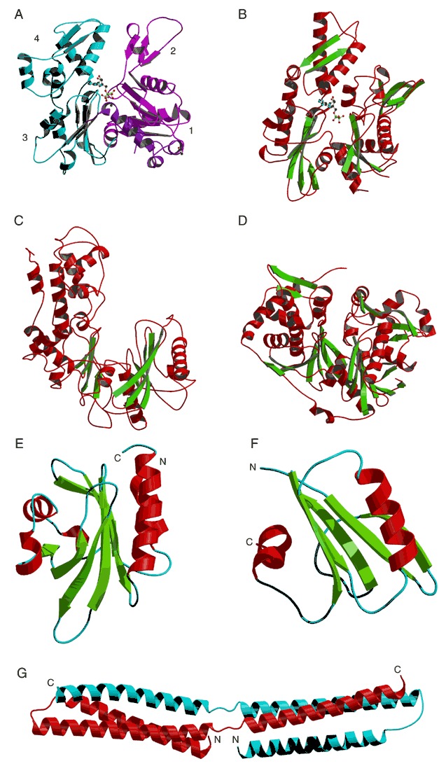
Structures containing the actin fold (A to D) and structures that bind to actin (E to G). (A) Actin complexed with ATP (PDB entry 1atn). Domains 1 and 2 are colored purple and blue, respectively. Subdomains 1 to 4 are labeled. ATP is shown as a ball‐and‐stick model. (B) The N‐terminal fragment of heat‐shock cognate protein (hsc70) complexed with ADP (PDB entry 1nga). ADP is shown as a ball‐and‐stick model. (C) Hexokinase B in its open form (PDB entry 2yhx). Structures of (D) glycerol kinase (PDB entry 1gla), (E) profilin (PDB entry 2btf), and (F) severin (NMR structure, PDB entry 1svq). (G) The spectrin repeat is shown as a dimer (PDB entry 1spc), with the two monomers colored red and blue, respectively.