
Figure 33.
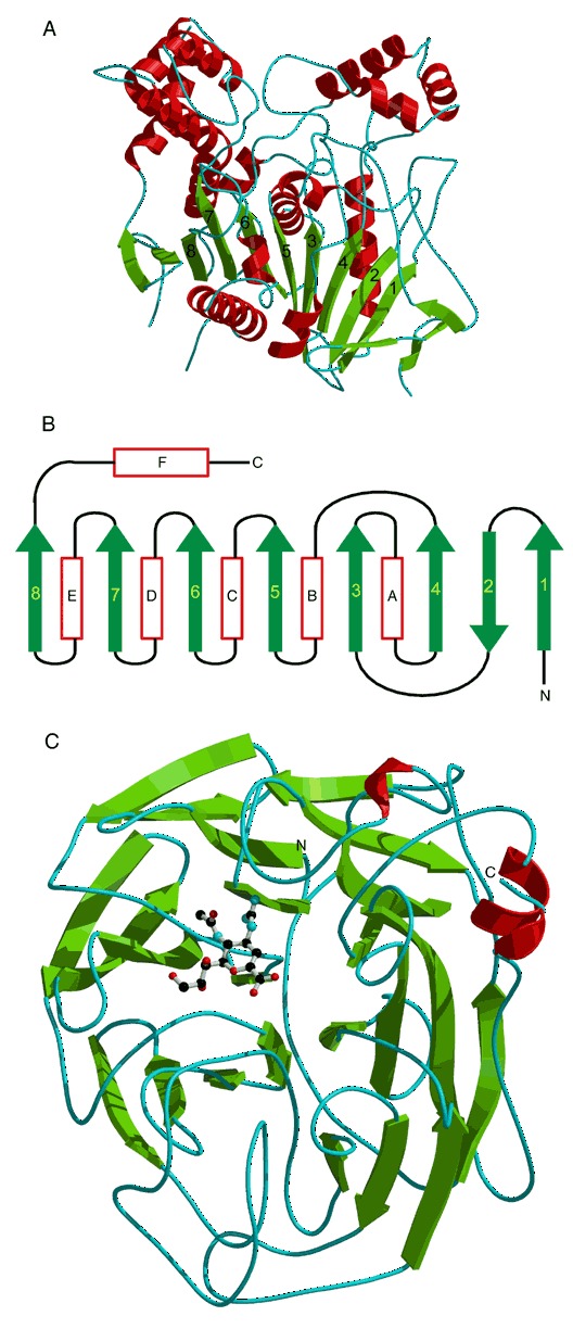
(A) Structure of acetylcholinesterase from Torpedo Californica. The central eight‐stranded β‐sheet is shown in green. (PDB entry 1ack). (B) Diagram of the secondary structure topology common to all α/β hydrolases. The strands are numbered from one to eight and helices are labeled A through F. (C) Structure of an influenza virus neuraminidase complexed with an inhibitor (PDB entry 1nnc). The active site‐bound inhibitor is shown as ball‐and‐stick model.