
Figure 35.
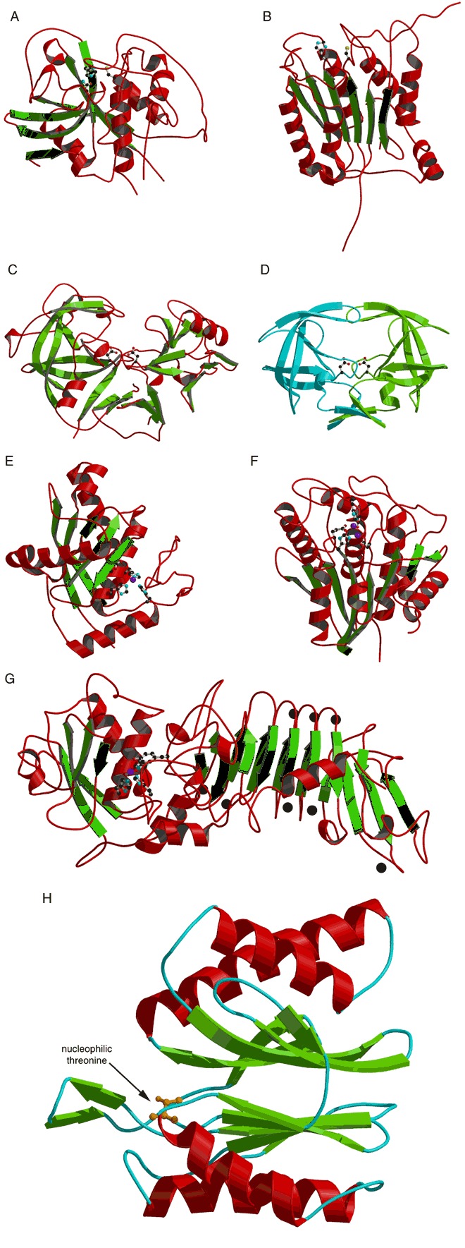
Examples of cysteine proteases (A and B), aspartic proteases (C and D), and metalloproteases (E to G). The side chains of critical active site residues mentioned in the text are shown as ball‐and‐stick models. Zinc atoms are shown as magenta balls. (A)The papain‐like cysteine protease, human cathepsin B (PDB entry: 1huc). (B) Interleukin‐1b converting enzyme (ICE; PDB entry: 1ice). (C) Pepsin‐like protease, human renin (PDB entry 1bbs). (D) The retroviral protease, HIV‐1 protease (PDB 1hpx). (E) Structure of a zinc‐dependent endopeptidase, a metzincin, snake venom adamalysin II (PDB entry 1iag). (F) Structure of a zinc‐dependent exopeptidase, aminopeptidase from Aeromonas proteolytica (PDB entry 1amp). (G) Structure of alkaline protease, a metzincin from Pseudomonas aeruginosa (PDB entry 1akl). The active site zinc and coordinated side chains are shown as ball‐and‐stick models in the N‐terminal catalytic domain (left side). Bound calcium ions are shown as black balls in the C‐terminal parallel β‐roll domain (right side). (H) A catalytic β‐subunit from the 20S yeast proteasome as a representative of the Ntn fold (PDB entry 1ryp). The nucleophilic threonine is shown as a ball‐and‐stick model.