
Figure 36.
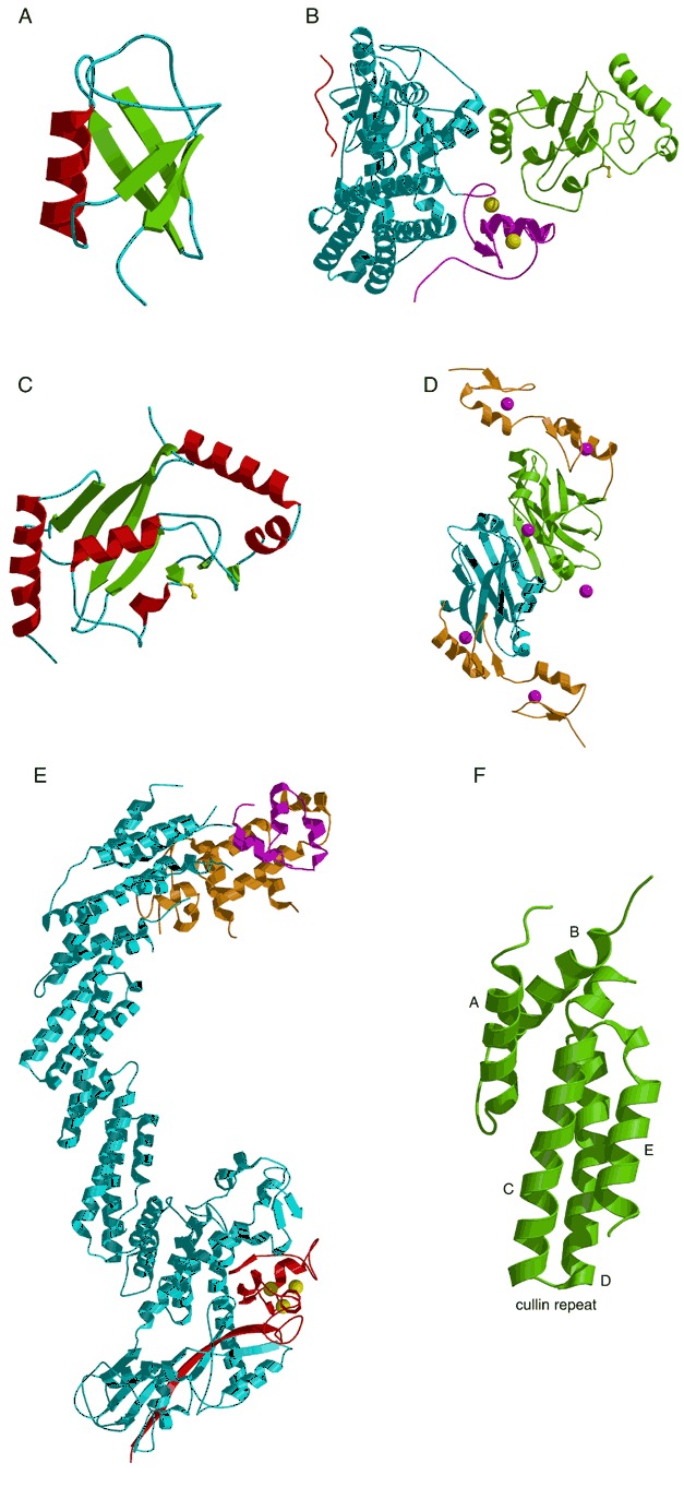
Enzymes of the ubiquitin pathway. (A) The structure of ubiquitin (PDB entry 1ubq). (B) Complex between the ubiquitin‐conjugating E3 enzyme c‐Cbl (cyan) and the E2 enzyme UbcH7 (green; PDB code 1fbv). The c‐Cbl RING domain is colored magenta with Zn shown as yellow balls and the c‐Cbl‐bound ZAP‐70 peptide is shown in red. The active site Cys86 of UbcH7 is shown as a yellow ball‐and‐stick model. (C) SUMO E2 enzyme Ubc9 with active site Cys93 shown as a yellow ball‐and‐stick model (PDB entry 1kps). (D) Siah homodimer (PDB entry 1k2f). The monomers are colored blue and green, respectively. The RING domains are in orange. Zinc atoms are magenta spheres. (E) Cul1‐Rbx1‐Skp1‐F‐box‐Skp2 complex. The Cul1, Ring‐Box, SKP1, and Skp2‐F‐box are colored in cyan, red, orange, and magenta, respectively (PDB code 1ldk). Zn ions are shown as yellow spheres. (F) The second cullin repeat from cul1 (PDB entry 1ldk).