
Figure 37.
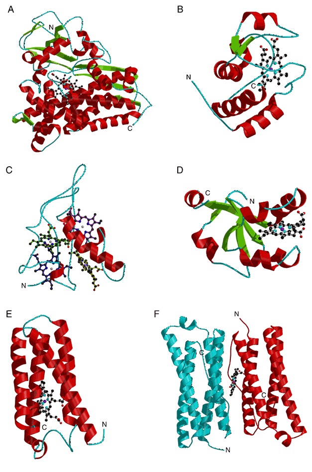
Structures showing the fold of (A) cytochrome P450‐CAM (PDB entry 1phc), (B) cytochrome c (PDB entry 1ycc), (C) cytochrome c3 (PDB entry 2cy3) with the four heme molecules colored differently, (D) cytochrome b5 (PDB entry 1cyo), (E) cytochrome b562 (PDB entry 1cgn), and (F) a dimer of bacterioferritin (also known as cytochrome b1; PDB entry 1bcf). The heme molecules are shown by ball‐and‐stick models.