
Figure 38.
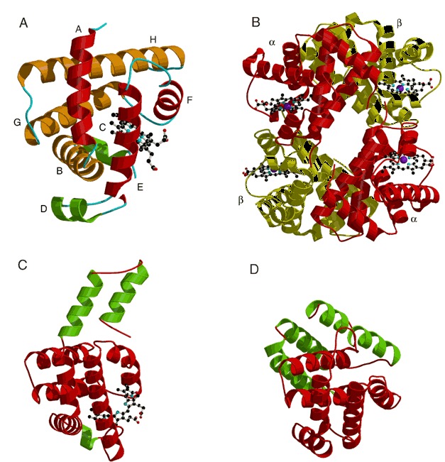
Structures of globin‐like proteins from each of the three globin‐like families. All three proteins are shown in the same orientation with respect to the globin fold. Helices that are not considered part of the core globin fold are colored green. (A) Sperm whale myoglobin (PDB entry 1mbd). Helices are labeled A to H according to traditional globin nomenclature. Helices A, E, and F are colored red, while helices B, G, and H are tan. The heme group is represented by a ball‐and‐stick model. (B) Structure of human deoxyhemoglobin showing all four subunits (PDB entry 1hhb). The heme groups are represented by ball‐and‐stick models. The α subunits are colored red and the β subunits are colored yellow. (C) C‐phycocyanin from cyanobacteria (PDB entry 1cpc). The phycocyanobilin cofactor is represented by a ball‐and‐stick model. (D) Colicin A from E. coli (PDB entry 1col).