
Figure 39.
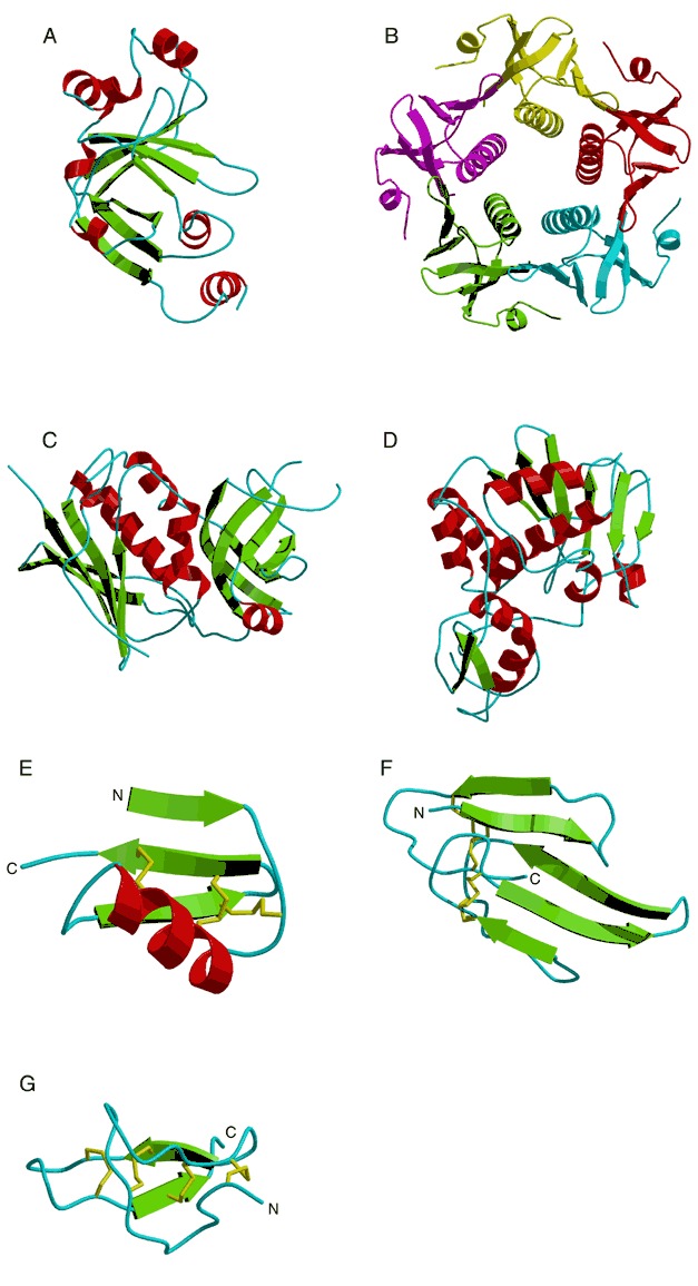
Tertiary folds of toxins. (A) ADP ribosylation domain of diphtheria toxin (PDB entry 1ddt). (B) Pentameric B domain of bacteria AB5 cholera toxin with each domain colored differently (PDB entry 1chp). (C) Superantigen toxin (PDB entry 1se2). (D) Ricin A chain (PDB entry 1rtc) The structure of (E) scorpion toxin (PDB entry 1mtx), (F) snake toxin (PDB entry 1nxb), and (G) spider toxin (PDB entry 1eit). Disulfide bonds are represented by yellow stick models.