
Figure 41.
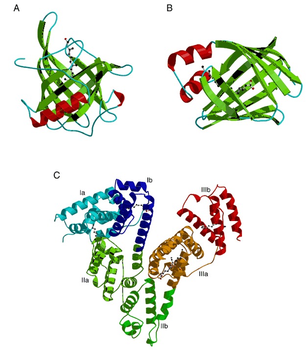
Lipid‐binding proteins with respective ligands shown as ball‐and‐stick models. (A) Human retinol‐binding protein bound to retinol (PDB entry 1rbp), an example of a lipocalin. (B) Rat intestinal fatty acid‐binding protein bound to palmitate (PDB entry 2ifb). (C) Human serum albumin with five bound myristate molecules (PDB entry 1bj5). Note that a helix spans both the I/II and II/III domain boundaries. This results in 28 instead of 30 helices in the structure.