
Figure 42.
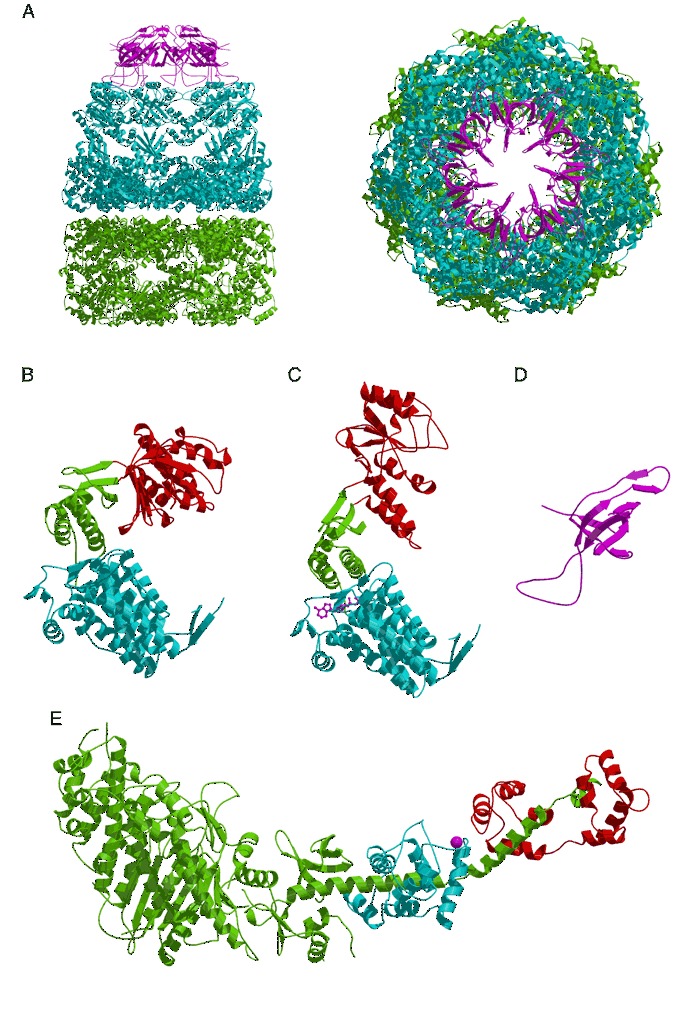
Chaperonin and myosin structures. (A) The GroEL/GroES complex viewed from the side (left) and top (right; PDB entry 1aon). The GroEL trans ring is green, the cis ring is blue, and the GroES ring is magenta. The domain structure of GroEL in the (B) trans and (C) cis rings of the GroEL/GroES complex (PDB entry 1aon). The equatorial domains are blue, the intermediate domains are green, and the apical domains are red. ADP is shown as a ball‐and‐stick model. (D) The GroES subunit (PDB entry 1aon). (E) The structure of the scallop myosin subfragment S1 (PDB entry 1b7t). Bound Ca2+ is shown as a small purple sphere. The heavy‐chain domain is shown in green. The essential and regulatory light chains are colored blue and red, respectively.