
Figure 47.
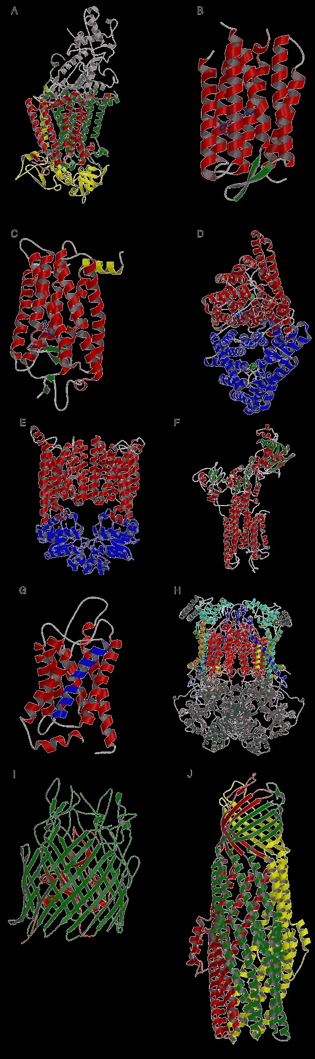
Structures of integral membrane proteins. (A) Photosynthetic reaction center (PDB entry 1prc). The four protein subunits are shown: cytochrome (gray), M (green), L (red), and H (yellow). (B) The structure of Halobacterium salinarum bacteriorhodopsin (PDB entry 1c3w), with the cytoplasmic face at the top. The retinal chromophore is shown as a blue ball‐and‐stick model. (C) Rhodopsin from the outer segment of bovine rod photoreceptor cells (PDB entry 1f88). The receptor is shown with the C‐terminal (cytoplasmic) domain at the top, and the N‐terminal (extracellular) domain at the bottom of the diagram. The covalently linked eleven‐cis‐retinal ligand is shown as a blue ball‐and‐stick model. The additional eighth helix is colored in yellow. (D) The ClC chloride channel from Salmonella typhimurium (PDB entry 1kpl). The double barrel of the homodimer is shown with Cl− ions, visible in the pores, displayed as green spheres. View is perpendicular to the plane of the membrane with the two subunits colored red and blue. (E) The E. coli vitamin B12 transporter BtuCD. The dimer is depicted, with BtuC subunits in red and BtuD subunits in blue (PDB entry 1l7v). (F) Calcium ATPase from skeletal‐muscle sarcoplasmic reticulum in the E1 state with bound calcium ions displayed as orange spheres. The three cytoplasmic domains are at the top (PDB entry 1eul). (G) Bovine AQP1 water channel monomer (PDB entry 1j4n). View is from the plane of the membrane with the extracellular face at the top. The two membrane‐inserted helices that do not span the membrane are colored in blue. (H) Bovine cytochrome bc1complex (PDB entry 1bgy). The subunits with membrane‐spanning portions are colored in the following scheme. Cytochrome b is red, cytochrome c1 is aqua, Rieske iron‐sulfur protein is blue, subunit 7 is orange, subunit 10 is green, and subunit 11 is yellow. (I) The E. coli ferric enterobactin receptor FepA (PDB entry 1fep). The β‐barrel domain is shown in green and the N‐terminal plug domain is shown in red. (J) TolC outer membrane protein of E. coli (PDB entry 1ek9). The trimer is shown with the subunits colored separately.