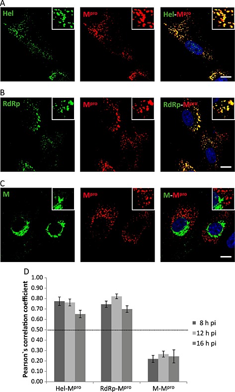
Figure 4.

Colocalization analysis of the BEV proteins involved in the replication/transcription processes.
A–C. E.Derm cells infected with BEV were fixed with 4% PFA in PBS at 8 h pi and used for dual immunolabelling with the anti‐Mpro antibody produced in rats (red) in combination with (A) rabbit anti‐Hel, (B) rabbit anti‐RdRp or (C) rabbit anti‐M (green). The nuclei were stained with DAPI (blue). The boxed areas are representative fields that are shown at higher magnification. Scale bars, 10 µm.
D. Quantitative analysis of the colocalization degree. The same experiment as described in A–C was also performed at 12 and 16 h pi, and Pearson's correlation coefficient was calculated for each pair of antibodies at the three analysed pi times, using for the analysis images (n = 5) containing an average of 20 cells each. Only the values above 0.5 were considered as indicative of colocalization.
