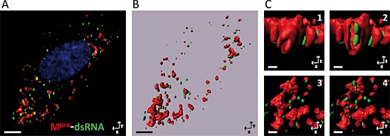
Figure 7.

Three‐dimensional reconstruction from images obtained by confocal microscopy: Mpro‐dsRNA. A series of consecutive images with a z‐step optimized were taken by confocal microscopy from E.Derm cells infected with BEV, fixed at 8 h pi and immunostained with the anti‐Mpro (red) and anti‐dsRNA (green) antibodies as described in the legend to Fig. 5A. The nuclei were stained with DAPI (blue).
A. Projection in the z‐axis of the captured images of an individual cell.
B. General view of the 3D reconstruction generated from the confocal images using the imaris software (Bitplane AG). Scale bars, 5 µm.
C. Enlarged area from the 3D reconstruction. (C1) Frontal view. (C2) Frontal view of a vertical section that shows the dsRNA inside the structure labelled with anti‐Mpro antibody. (C3) Top view. (C4) Top view of a cross section. Scale bars, 0.5 µm.
