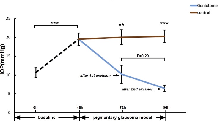
Fig 2. IOP reduction after TM excision.
After 48h perfusion with pigment, IOP in PG was significantly higher compared to baseline (n = 15, ***P<0.001). Twenty-four hours after a 90° excision of TM (G), IOP in G (n = 7) was lower than in C (n = 8, **P<0.01). Twenty-four hours after a second, adjacent excision of 90° TM, IOP in G appeared to slightly reduce further (n = 7, P>0.05) remaining lower than C (n = 8, ***P<0.001).

