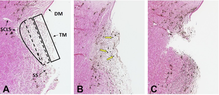
Fig 4. Histology.
Normal TM was lightly pigmented and consisted of three areas with different compactness and cell densities: the uveal meshwork (box with solid line, A), the corneoscleral meshwork (box with dashed line, Fig 4A), and the juxtacanalicular meshwork (solid line, A) adjacent to small canal-like segments characteristic of the porcine angular aqueous plexus (SCLS). Pigment granules were observed in all layers of TM (B, yellow arrows) after perfusion with pigment-supplemented media. After TM excision, a large, full-thickness portion of TM was removed (C). Trabecular meshwork: TM, Schlemm’s canal-like segments (SCLS) of the porcine angular aqueous plexus, scleral spur (SS), Descemet's membrane (DM).

