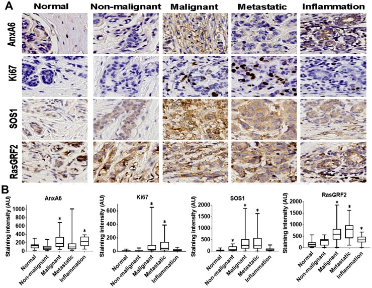
Fig 2. Expression of AnxA6 and proliferation markers in normal breast and breast disease tissues.
A) Thin sections of formalin-fixed paraffin-embedded tissues in a broad-spectrum breast disease TMA were stained with antibodies against the indicated proteins. Shown are representative stained tissues. B) The stained tissues were digitally scanned and digitally analyzed using the Tissue IA software. Shown are boxplots depicting the mean staining intensity and distribution of AnxA6, Ki67, SOS1 and GRF2 staining intensity in the normal and the variety of breast disease tissues. * indicates p<0.05 for relative staining intensity of each protein in the indicated subgroup of breast disease tissues compared to normal breast tissues.

