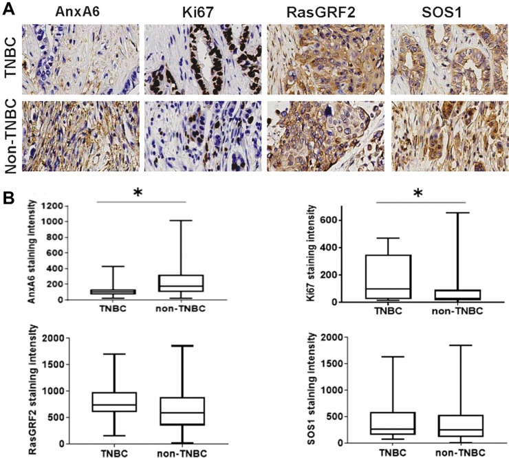
Fig 3. Expression of AnxA6 and proliferation markers in malignant and metastasis breast cancer tissues.
Tissues were processed as described in Fig 2 and the malignant and metastasis tissues stratified into TNBC and non-TNBC subsets. Shown are representative stained tumor tissues (A) and analysis of the staining intensities of the indicated proteins by using the Tissues IA software (B). * indicates p<0.05 for the mean staining intensity of each protein in the TNBC subset compared to the non-TNBC subset.

