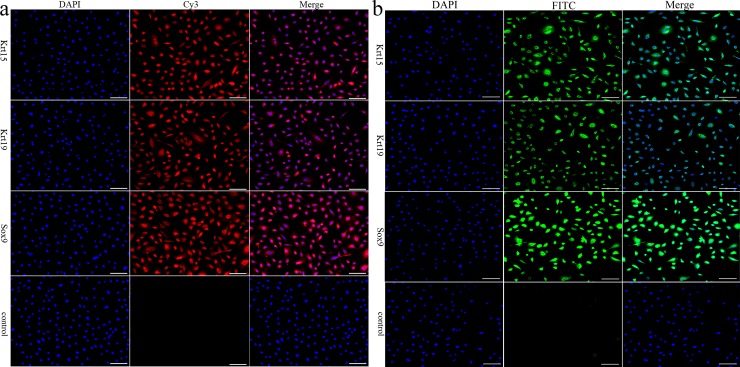
Fig 2. Identification of the in vitro cultured HFSCs.
(a): Immunocytochemistry staining of Krt15, Krt19 and Sox9 in the anagen secondary HFSCs. (b): Immunocytochemistry staining of Krt15, Krt19 and Sox9 in the telogen phase secondary HFSCs. Scale bars 100 μm. Control was HFSCs incubated with 10% goat serum instead of primary antibody; red, Cy3–conjugated goat anti-rabbit IgG; green, Fluorescein (FITC)–conjugated goat anti-rabbit IgG; blue, DAPI staining.

