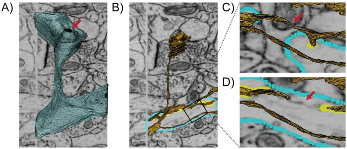
Fig 2. Initial surface mesh model of a single spine scene with subcellular organelles imaged by Focused-ion Beam Milling Scanning Electron Microscopy (FIB-SEM) (overlaid), courtesy of Wu et al. [7], contains many mesh artifacts and is not compatible with physics-based simulations.
A) The blue surface represents the Plasma Membrane (PM) which contains a hole indicated by the red arrow. B) The yellow surface represents the membrane of the Endoplasmic Reticulum (ER). C, D) are two views rotated and zoomed-in on B showing a disconnected region of the ER proofed against micrographs taken at different z-axis locations. An untraced ghost, indicated by the red arrow, appears between the disconnected segments which suggests a possible error in the segmentation.

