Figure 4.

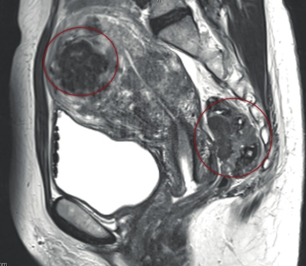
MRI picture of the pelvis. DE in the rectum with small, dense adhesions between the posterior wall of the cervix and ante- rior wall of the rectum (right circle) and adenomyosis (left circle).

MRI picture of the pelvis. DE in the rectum with small, dense adhesions between the posterior wall of the cervix and ante- rior wall of the rectum (right circle) and adenomyosis (left circle).