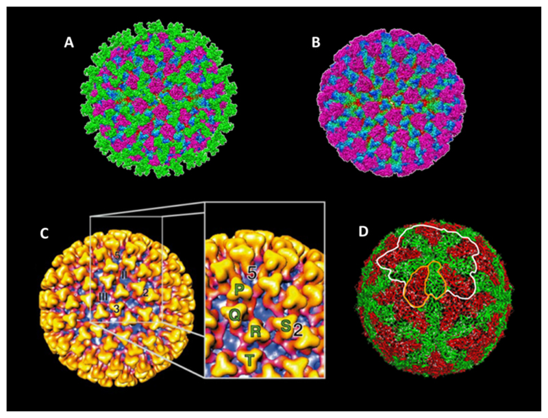
Fig. 2. Structure of the particle and inner capsid layers.
(A) The 7 A ° Cryo-EM structure of the complete particle. Triskelion shaped trimers of VP2 (green) are exposed on the outer surface. VP5 trimers (purple) sit under VP2 trimers between the triskelion arms. Regions of the underlying VP7 (blue to green gradient) and VP3 (red) can be seen. (Adopted from Zhang et al., 2010.) (B) The structure of the mature particle lacking the VP2 trimer layer (colouring as in A). (Adopted from Zhang et al., 2010 (C)). The 23 A ° Cryo-EM structure of the core particle. VP7 trimers (gold to red gradient) are surface exposed with regions of the VP3 sub-core layer (purple) visible below. 2, 3 and 5 denote the icosahedral 2-fold, 3-fold and 5-fold symmetry axis respectively. II and III denote the position of class II and class III channels. The cut away annotates the P, Q, R, S and T crystallographic positions. (Adapted from Grimes et al., 1997 (D)). The 3.5 A ° X-ray crystal structure of the sub-core particle. Two isoforms of VP3 are present, A (green) and B (red). These form a dimer (outlined in orange) which associate to form a decamer (outlined in white), twelve of which compose the sub-core layer. (Adapted from Grimes et al., 1998).

