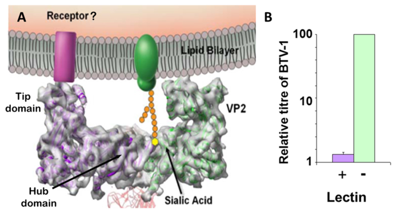
Fig. 4. BTV cell attachment.
(A) The model of cell surface interaction of VP2. The side on profile of a VP2 triskelion trimer is shown. Two VP2 molecules can be seen (green and purple) attached to an underlying VP7 trimer (red). Note the L shaped profile of the trimer. The hub domain binds a sialidated cell surface protein (green) in a receptor pocket. The tip domain may interact with an additional co-receptor (purple). (Adapted from Zhang et al., 2010). (B) Sialic acid interaction is necessary but not sufficient for cell entry. Pre-treatment of HeLa cells with wheat germ agglutinin (lectin) drastically decreases but does not inhibit the replication of BTV.

