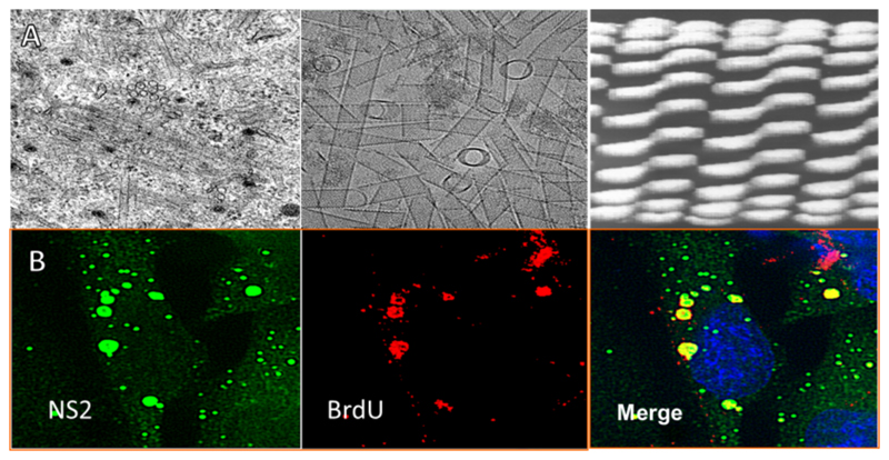
Fig. 5. Cellular morphogenesis by non-structural proteins.
(A) NS1 forms tubules. Transmission electron micrographs show the morphology of NS1. The left panel shows BSR cells 24 hrs post BTV infection, tubular structures are clearly visible in the cytosol. The middle panel displays purified NS1 tubules expressed using a recombinant baculovirus system. The right panel shows the 40 Å Cryo-EM structure of NS1 tubules, which illustrate a coiled tube of dimers. (B) NS2 forms viral inclusion bodies. The left panel shows cells 17 hrs post infection stained for NS2 (green). The middle panel displays the same cells stained for bromo-deoxyuridine (BrDU) (red). The right panel represents the merged image. Globular structures with peri-nuclear localisation are seen with the co-localisation of BrDU staining. (Adapted from Kar et al., 2007).

