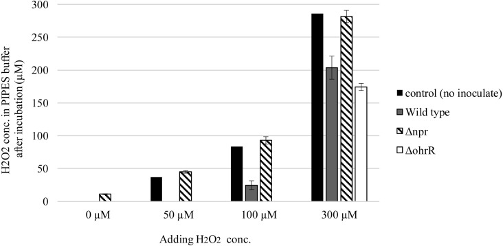
Fig. 4.
H2O2 concentration of wild type and Δnpr in PIPES buffer.
Strains precultured overnight at 37°C were inoculated into fresh MRS medium at a final OD600 of 0.05. The cells were used after static culture at 37°C for 5 hr. They were washed twice with PIPES buffer (pH 6.8) and resuspended in 10 mL H2O2 adjusted to 0 to 300 µM with PIPES buffer. After incubation at 37°C for 2 hr, the cells were harvested by centrifugation (10,000 g, 3 min). Twenty microliters of the supernatant was used for measuring the H2O2 concentration. After measuring the wavelength at 727 nm, the chromogenic reagent DA64 was used to quantify H2O2 based on the standard curve. The data are shown as the mean ± SE of three independent experiments.

