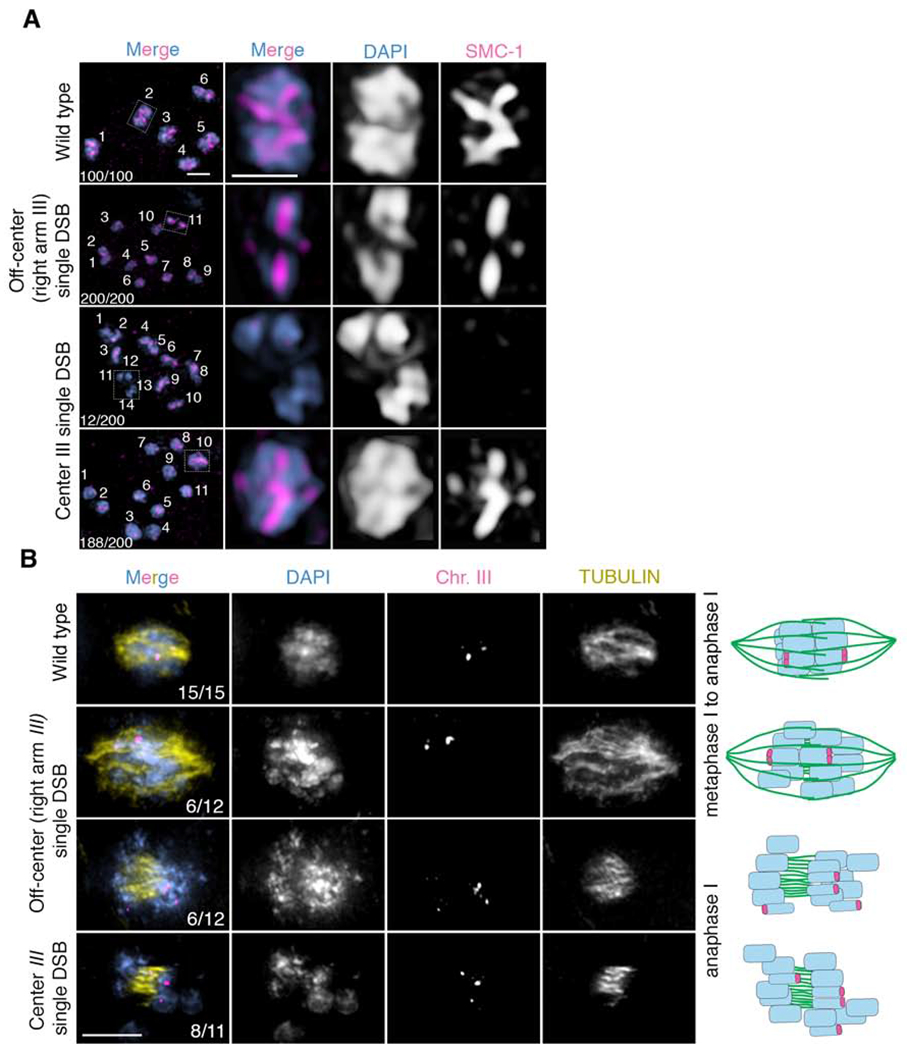
Figure 2. A single centered crossover results in premature sister separation.

(A) Immunolocalization of SMC-1 (red) and DAPI (blue) in diakinesis stage oocytes in wild type, spo-11 mutants with a single Mos1 insertion at an off-center position (right arm) on chromosome III, and spo-11 mutants with a single Mos1 insertion at the center of chromosome III after heat shock, all of which underwent emb-30 depletion by RNAi to induce arrest of oocytes before the meiotic division to facilitate this analysis. 12 out of 200 oocytes in spo-11 mutants with a single Mos1 insertion at the center of chromosome III showed premature separation of sister chromatids after heat shock as evidenced by the presence of 14 DAPI-stained bodies (10 univalents + 4 separated sister chromatids) and absence of SMC-1 staining in 4 DAPI-stained bodies (presumably the 4 prematurely separated sister chromatids). (B) Immunofluorescence-FISH images of metaphase to anaphase I nuclei. On the first row are representative images of wild-type nuclei at the metaphase to anaphase I transition with a chromosome III FISH probe (magenta) and tubulin (yellow). The two FISH signals (foci) show separating homologs but joined sister chromatids (n=15/15 nuclei examined). The second and third rows are representative images of metaphase to anaphase I and anaphase I nuclei, respectively, from worms harboring a single Mos1 insertion at an off-center position (right arm) on chromosome III. Both nuclei with separating homologs but joined sister chromatids (n=6/12), as seen in wild type, and nuclei with premature sister separation (n=6/12), as depicted by more than two FISH signals, were detected. The last row depicts anaphase I for worms with a single Mos1 insertion at the center of chromosome III. Sister chromatid separation is evidenced by the presence of more than two FISH signals (n=8/11). Numbers in the first column represent number observed/total number scored. Bar, 2 μm.
