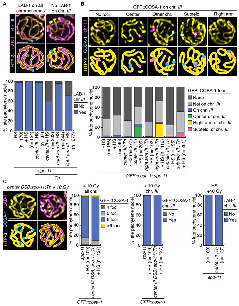
Figure 3. Chromosome remodeling defects are already observed at late pachytene stage and can be rescued by additional exogenous DSBs.

(A) Immunolocalization of LAB-1 in late pachytene on chromosome III following heat shock-induced DSB formation. HTP-3 (yellow), LAB-1 (magenta) and a FISH probe for chromosome III (blue) are shown. Illustrations for each observed localization pattern are shown in the lower panel. Histogram shows the quantification of various LAB-1 localization patterns in late pachytene nuclei subjected to heat shock-induced DSBs at different locations on chromosome III. (B) Crossover precursor marker COSA-1 localizes at high frequency to Mos1-induced CO sites. Late pachytene nuclei of whole mounts hybridized with a FISH probe recognizing chromosome III (magenta) showing the localization of chromosome axis marker HTP-3 (yellow) and GFP::COSA-1 (blue). Illustrations depict the different localizations observed for COSA-1. Histogram shows the quantification of COSA-1 foci in late pachytene nuclei from lines with DSBs induced at the indicated positions on chromosome III (identified by FISH). Similar to other studies, GFP::COSA-1 foci were also detected on chromosomes in spo-11 mutants, potentially reflecting spontaneous DNA lesions [43, 44]. n, number of late pachytene nuclei scored; HS, heat shock. (C) Exogenous DSB formation by γ-IR rescues CO formation and LAB-1 localization defects in late pachytene nuclei subjected to a single Mos1-induced DSB at the center of chromosome III. HTP-3 (yellow), COSA-1 (blue) and LAB-1 (magenta). Histograms show quantifications of COSA-1 foci and LAB-1 localization pattern on chromosome III (identified by FISH). Bars, 2 μm. See also Figure S3 and Data S3, S4 and S5.
