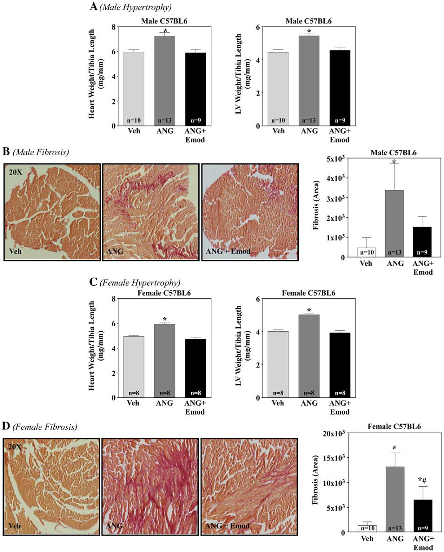
Figure 6. Emodin attenuated angiotensin II-induced pathological hypertrophy and fibrosis in male and female mice.

C57BL/6 male and female mice were surgically implanted with a sham (Veh) or micro-osmotic pump containing angiotensin II (Ang; 1.5 μg/kg/min) and dosed with or without emodin (Emod, 30 mg/kg/day) for 14 days. 14 days post Ang, whole hearts and left ventricles were dissected and assessed for hypertrophy and fibrosis. Whole heart weight to tibia length (HW/TL, mg/mm) and left ventricle weight to tibia length (LV/TL, mg/mm) of sham (Veh), Ang and Ang+Emod treated C57BL/6 male (A) and female (C) mice were examined at study end (14 days). Left ventriclular collagen of sham (Veh), Ang and Ang+Emod treated C57BL/6 mice was assessed via PicroSirius Red staining for male (B) and female (D) mice. ImageJ software was used to determine fibrotic area. GraphPad Prism was used to examine statistical significance. One-way ANOVA with Tukey’s post-hoc analysis was used. Significance was set at p<0.05.
