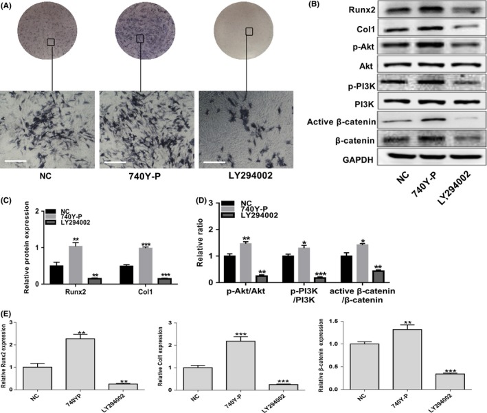
FIGURE 4.

The PI3K/Akt/β‐catenin pathway positively regulates osteogenic differentiation in EMSCs. Wild‐type (NC) EMSCs were treated with the PI3K/Akt signalling PI3K agonist 740Y‐P and inhibitor LY294002 during cultured in osteogenic induction medium for 7 d. (A) ALP staining intensity was observed by optical microscopy. Scale bar represents 50 μm. (B) The protein levels of Runx2, Col1, PI3K, p‐PI3K, Akt, p‐Akt, β‐catenin and active β‐catenin were detected by Western blot analysis. Grayscale analysis was performed, and (C) the levels of Runx2 and Col1 proteins were expressed relative to the levels of GAPDH. (D) Phosphorylation of PI3K and Akt and activation of β‐catenin were analysed, and the results were represented as fraction of the control. (E) The mRNA levels of Runx2, Col1 and β‐catenin were examined by real‐time PCR normalized to GAPDH. Data are shown as mean ± SD from three independent experiments. **P < .01, **P < .01, ***P < .001
