Figure 6.

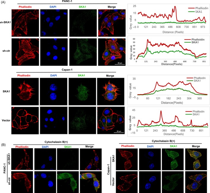
SKA1 modulates the formation of cytoskeletal actin filament and shared partial co‐localization with F‐actin in PDAC cells. A, Combined rhodamine‐phalloidin staining for F‐actin (red) and SKA1 (green) immunofluorescence, nuclei were counterstained with DAPI (blue). Upper two panels: stable knock‐down of SKA1 reduced actin protrusions and stress fibres in PANC‐1 cells. Lower two panels: stable overexpression of SKA1 altered the cellular morphology and lead to longer invadopodia formation compared with vector group in Capan‐1 cells. Additionally, SKA1 and phalloidin‐stained F‐actin shared partial co‐localization to the cytoplasm. Co‐localization correlation of intensity distributions curves between two channels was drawn by ImageJ and measured by Pearson's correlation coefficient analysis (right). B, With cytochalasin B treatment, F‐actin filaments stained with phalloidin were not seen in both PANC‐1 and Capan‐1 cells’ infectants
