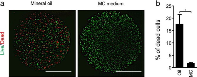
Figure 4.
Viability of encapsulated Hep G2 cells aggregated by conventional oil and MC medium methods. (a) ECM gel capsules were stained with propidium iodide (red) to identify dead cells and calcein-AM (green) to identify living cells. (b) The percentage of dead cells in ECM capsules was calculated. ECM capsules treated with 4% paraformaldehyde were used as 100% dead spheroid controls. Bar = 500 µm. The data are the means ± SEMs, n = 3. *p < 0.05 by Student’s t-test.

