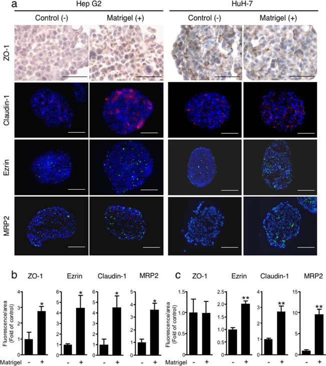
Figure 7.
Immunostaining of cell polarity-related proteins. (a) Control spheroids and ECM-loaded spheroids with 0.3 mg/ml Matrigel were fixed two days after injection into MC medium, followed by sectioning and staining for the tight junction-associated proteins ZO-1 (DAB staining) and Claudin-1, the apical marker Ezrin, the bile acid transporter MRP2 (red fluorescence) and staining with Hoechst 33342 for nuclei (blue). Bar = 50 µm. Positive areas for ZO-1, Claudin-1, Ezrin, and MRP2 were quantified and normalized to the nuclear area of Hep G2 (b) and HuH-7 (c) spheroids. The data are shown as the fold of expression in control spheroids. The data are the means ± SEMs, n = 3–4. *p < 0.05. **p < 0.01 by Student t-test.

