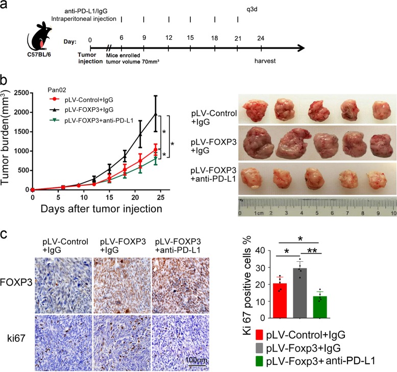
Fig. 4.
Anti-PD-L1 antibody counteracts the growth of PDAC induced by C-FOXP3 in vivo. a C57BL/6 mice were inoculated subcutaneously with murine pancreatic tumor cells in their right flanks (5 mice/group). When tumors reached approximately 70 mm3 in size, mice were treated with isotype control or anti-PD-L1 for 3 weeks. b Mean (left) and individual (right) tumor volumes over time. On day 24, tumor growth was evaluated by measuring tumor volumes and compared statistically by one-way ANOVA with the Bonferroni post hoc test. (*p < 0.05). Line chart: points: mean, bars: standard deviation, n = 5. c (Left) Immunohistochemical staining of FOXP3 and Ki67 in tumor slices. Representative images are shown, magnification: ×200. Ki67 (right) expression levels for the slides are summarized in the graph. Histogram (columns: mean, bars: standard deviation, n = 5), p values were calculated by one-way ANOVA tests, *p < 0.05, **p < 0.01

