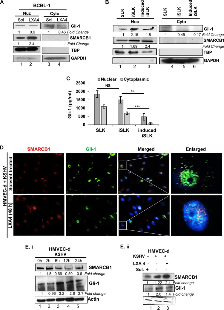
FIG 6.
LXA4 treatment regulates the nuclear translocation of Gli-1 and the SMARCB1–Gli-1 interaction in the nuclei of KSHV-infected cells. (A) The nuclear (Nuc) and cytoplasmic (Cyto) fractions were isolated from BCBL-1 cells and LXA4-treated BCBL-1 using a nuclear isolation kit and immunoblotted with anti-SMARCB1 and anti-Gli-1 antibodies. These membranes were stripped and immunoblotted with anti-GAPDH and anti-TATA-binding protein (anti-TBP) antibodies to check the purity of the cytoplasmic and nuclear lysates, respectively, and to confirm equal loading. (B) Shuttling of Gli-1. Immunoblotting showed the total nuclear translocation of the Gli-1 protein in induced iSLK cells compared with that in iSLK cells. (C) Gli-1 expression in lysates obtained from the nuclear and cytoplasmic fractions of SLK cells, latently infected SLK cells (iSLK), and induced iSLK cells was estimated using an ELISA kit. The experiments were repeated three times, and the results are expressed as the mean ± SD. **, P < 0.01; ***, P < 0.001; NS, not significant. (D) Nuclear translocation of Gli-1 during the treatment of de novo KSHV-infected HMVEC-d cells with LXA4. (E) (i) HMVEC-d cells were infected with KSHV (30 DNA copies/cell) for 0, 2, 6, 12, and 24 h. The Western blot shows the fold change in protein expression for SMARCB1 and Gli-1 in KSHV-infected HMVEC-d cells when expression for uninfected cells was considered 1. (ii) HMVEC-d cells were infected with KSHV (30 DNA copies/cell), and lysates were prepared from LXA4-treated cells or cells mock treated with solvent and were immunoblotted for SMARCB1 and Gli-1. The expression for uninfected HMVEC-d cells was considered 1. The values presented are the averages of results from three independent experiments, and statistical analysis was done by using Student's t test.

