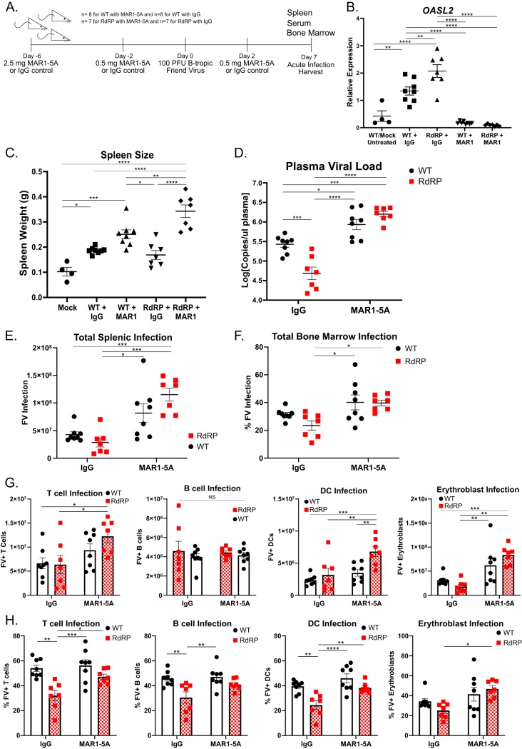
FIG 7.
Blockage of IFN receptor signaling eliminates protection against FV infection in RdRP mice. (A) Experimental design of anti-IFNAR-treated mice for infection with 100 PFU FV. Mixed-gender mice were treated with MAR1-5A or IgG control at indicated doses and time points pre- and postinfection. Mice were sacrificed at 7 dpi for evaluation of FV infection. For all experiments shown, n = 4 for mock (uninfected WT mice, included to show baseline spleen size), n = 8 for WT with MAR1-5A or IgG, and n = 7 for RdRP with MAR1-5A or IgG. (B) qRT-PCR from liver sections for the mRNA of the representative ISG Oasl2. (C) Spleen weights of mice described in panel A. (D) Plasma viral load of mice described in panel A. Data were log10 transformed prior to analysis. (E, G) Spleen cell analyses. Shown are total FV-positive (FV+) splenocytes (E) and FV+ T cells, B cells, DCs, and erythroblasts (G) in the spleens of infected mice as determined by flow cytometry. (F, H) Bone marrow cell analyses. Shown are percent FV-positive total cells (F) and percent FV-positive T cells, B cells, DCs, and erythroblasts (H) in the bone marrows of infected mice as determined by flow cytometry. A one-way ANOVA followed by a Tukey’s test to determine significance was used to analyze spleen weight and Oasl2 expression. All other data were analyzed using a two-way ANOVA followed by a Tukey’s test to determine significance. *, P < 0.05; **, P < 0.01; ***, P < 0.001; ****, P < 0.0001.

