FIG 3.
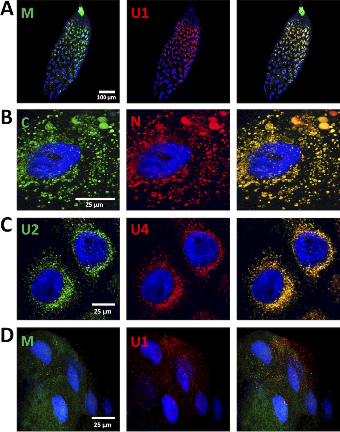
Colocalization of FBNSV segments in AMG and PSG. The colors of the fluorescent probes and the targeted segment pairs are indicated. Three additional pairs of segments were tested in the AMG: 32 aphids from four experiments for the pair M/U1 and 24 aphids from three experiments for the pairs C/N and U2/U4. Illustrative images are, shown, respectively, in panels A, B, and C. In the PSG, the additional segment pair M/U1 was observed in 6 aphids from three experiments, and a representative image is shown in panel D. Split-color channels are shown in the left and middle panels whereas merged images are shown in the right panel. All images correspond to maximum-intensity projections. Cell nuclei are stained with 4′,6′-diamidino-2-phenylindole (DAPI; blue).

