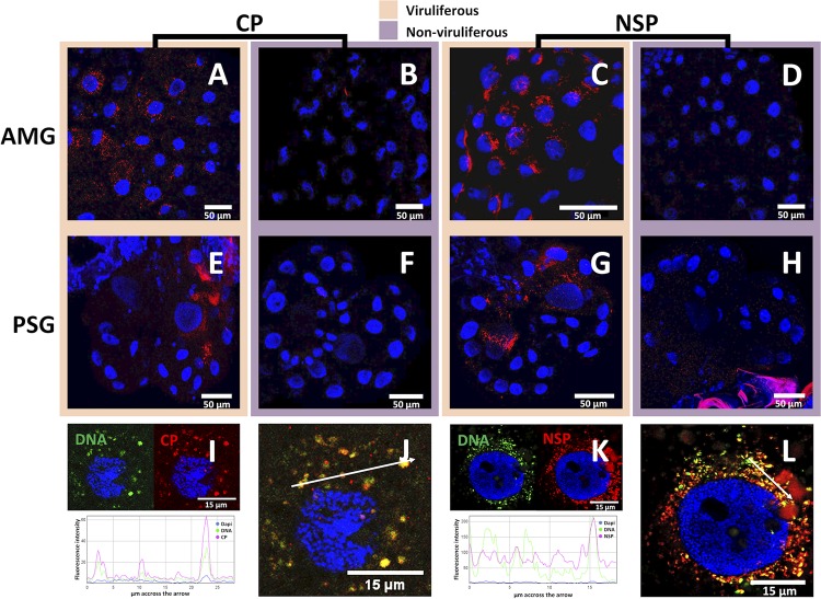
FIG 4.
Localization of FBNSV DNA, CP, and NSP in AMG and PSG of A. pisum. FBNSV CP is labeled by IF in the AMG (A and B) and PSG (E and F) of viruliferous (A and E) and nonviruliferous (B and F) aphids. NSP is labeled by IF in the AMG (C and D) and PSG (G and H) of viruliferous (C and G) and nonviruliferous (D and H) aphids. When viral DNA and either CP or NSP (FISH and IF) were colabeled in AMG, the DNA probe targeting all eight genome segments appears green whereas the specific CP or NSP antibody appears red (I to L). In each case, 30 aphids from three experiments were observed, and one representative image is shown. Split-color channel images in panels I and K correspond to the merged-color channel images in panels J and L, respectively. The graphics represent the colocalization profiles between either DNA (green curve) and CP (red curve) (I) or DNA (green curve) and NSP (red curve) (K). Fluorescence intensity was measured along the white arrows drawn in panels J and L. Images in panels A to H correspond to maximum-intensity projections, and images in panels I to L correspond to single optical sections. Cell nuclei are stained with 4′,6′-diamidino-2-phenylindole (DAPI; blue).

