FIG 5.
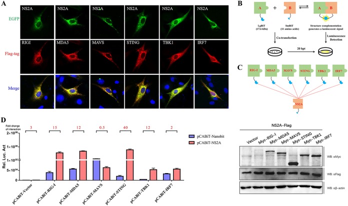
DTMUV NS2A inhibits RIG-I-, MAD5-, MAVS-, and STING-mediated IFN induction. (A) BHK-21 cells were cotransfected with each of the components (pCAGGS-RIG-I, MDA5, MAVS, STING, TBK1, and IRF7-Flag) (400 ng/well) and pCAGGS-NS2A-His (400 ng/well). At 24 h posttransfection, cells were washed with cold PBS three times and subsequently fixed in 4% paraformaldehyde overnight at 4°C for IFA. (B, C) Schematics of the NanoBiT assay and plasmid construction. (D) DEFs were cotransfected with pCABiT-NS2A-SmBiT-Flag (400 ng/well) and plasmids encoding each of the components (pCABiT-RIG-I, MDA5, MAVS, STING, TBK1, and IRF7-LgBiT-Myc) (400 ng/well), and the luciferase activities were measured at 20 h posttransfection. Luminescence was measured at a user-defined time point or continuously for up to 2 h. According to the operational instructions, luminescence 10-fold higher than that of the negative control indicated a specific PPI (protein-protein interaction). Protein expression levels were determined by Western blot analysis. All data are represented as the mean ± SEM (n = 4). Significant differences from the mock groups are indicated by *, P < 0.05; **, P < 0.01; ***, P < 0.001.

