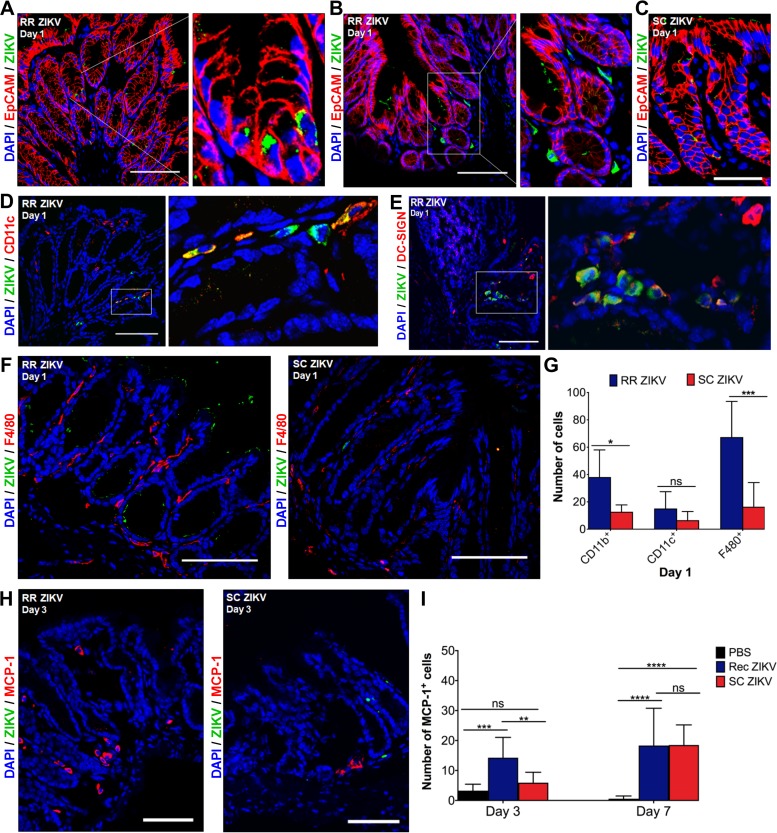
FIG 2.
ZIKV infects columnar epithelial cells and dendritic cells in the rectal mucosa 1 day postrectal inoculation. (A, B) Representative immunofluorescence images of rectal tissue from mice infected via the rectal route (RR) at 1 day postinoculation (dpi). Rectal tissue sections were immunostained for EpCAM (red) and ZIKV (green), and nuclei were stained with DAPI (blue). Inset in A shows a magnified view of infected epithelial cells comprising intestinal glands. Inset in B shows a magnified view of infected nonepithelial cells within the lamina propria. (C) Representative image of rectal tissue infected via the subcutaneous route (SC ZIKV) at 1 dpi. (D, E) Representative immunofluorescence images of rectal tissue for RR ZIKV at 1 dpi. Tissue was immunostained for ZIKV (green) and with anti-CD11c (red) or anti-DC-SIGN (red) at 1 dpi. Inset panels show ZIKV-infected CD11c+ or DC-SIGN+ dendritic cells (yellow) within the lamina propria. (F) Images of infected rectal tissue sections for RR and SC ZIKV at 1 dpi. Shown are ZIKV-infected cells (green) and F4/80+ macrophages (red). (G) Quantification of CD11b+, CD11c+, and F4/80+ cells in the rectal mucosa at 1 dpi. (H) MCP-1+ cells in rectal tissue from RR and SC ZIKV at 3 dpi. All images for A to F and H are representative of five different animals per group and were collected at ×40 magnification; scale bar, 50 μm. (I) Quantification of MCP-1+ cells for RR and SC ZIKV at 3 and 7 dpi. Multiple-comparison two-way analysis of variance (ANOVA) and Tukey tests were conducted (*, P < 0.05; **, P < 0.001; ***, P < 0.0001; ****, P < 0.00001; ns, no significance).

