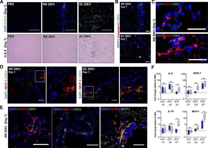
FIG 4.
Rectal ZIKV inoculation leads to reduced pathology and inflammation in the brain. (A, Top) Immunofluorescence images of uninfected (PBS) and infected brain tissue sections at 7 dpi (RR versus SC routes). Tissues were immunostained with anti-flavivirus antibody (green), and nuclei were stained with DAPI (blue). (A, Bottom) H&E images of brain tissue sections at 7 dpi (×10 objective). (B) Immunofluorescence images showing ZIKV infection (green) and cleaved caspase-3 immunostaining (red) of infected brain sections at 7 dpi. Images were collected at ×40 magnification; scale bar, 50 μm. (C) Images of ZIKV-infected CD31+ endothelial cells (top) and infected GFAP+ astrocytes (bottom) for RR ZIKV brain sections at 1 dpi. Images were collected at ×20 magnification; scale bar, 20 μm. (D) Immunofluorescence images showing ZIKV infection (green) and MCP-1+ cells (red) in RR or SC ZIKV-infected brains at 1 dpi. Images were collected at ×20 magnification; scale bar, 50 μm. (E) ZIKV-infected GFAP+ astrocytes (left), infected MCP-1+ cells (middle), and colocalization of MCP-1+ with GFAP+ cells (right) at 7 dpi. Images were collected at ×20 magnification; scale bar, 50 μm. (F) Fold change of IL-6, OAS-1, IL-10, and MCP-1 in the brain relative to the PBS control group at days 1, 3, and 7. Two-tailed unpaired, nonparametric Mann-Whitney tests were conducted (*, P < 0.05; **, P < 0.001; ns, no significance). Data are representative of one of two independent experiments.

