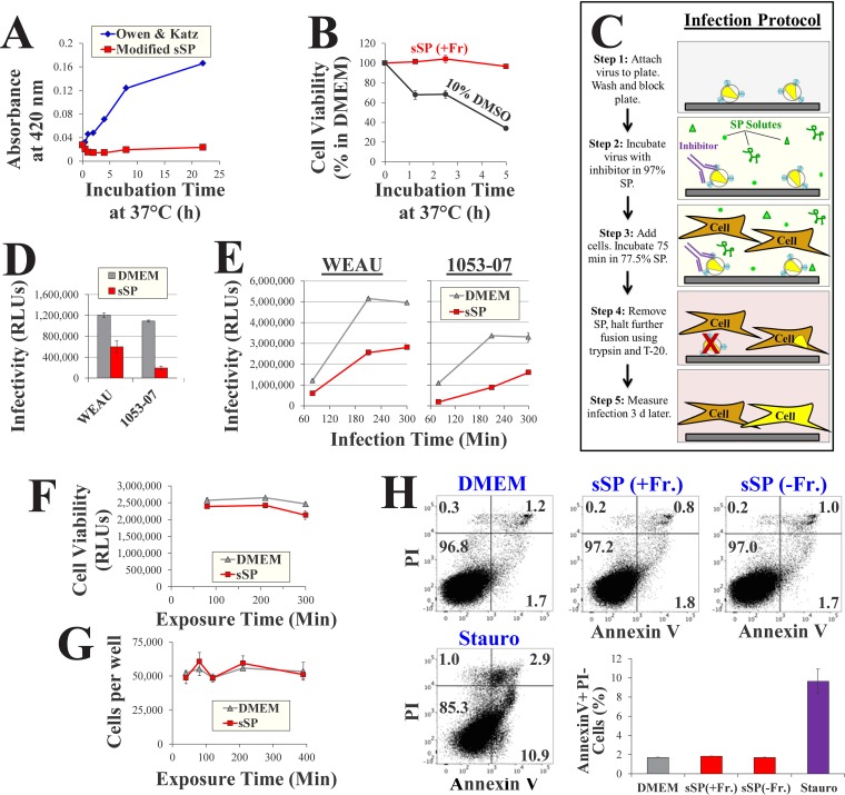
FIG 3.
HIV-1 infectivity and cell viability in a simulant of seminal plasma. (A) Precipitation in simulants of seminal plasma. Simulants were incubated at 37°C for different time periods, and precipitation was measured by determination of the absorbance at 420 nm. (B) Cell viability in sSP. Cf2Th CD4+ CCR5+ cells were incubated in sSP supplemented with 15 mM fructose [sSP(+Fr)] at 37°C for different time periods, washed, and further cultured in DMEM for 36 h. Cell viability was measured by an ATP-based assay. Samples exposed to the cytotoxic agent dimethyl sulfoxide (DMSO) were tested as a control. (C) Schematic of virus infectivity tests in SP. d, days. (D) Infection measured by the protocol outlined in panel C for viruses containing the indicated Envs. (E) Rate of HIV-1 entry in sSP. Cells were incubated with viruses in sSP or DMEM for different time periods before infection was halted by trypsin and T-20 (step 4). (F) Viability in samples treated as described in the legend to panel E was measured after 36 h. (G) Samples were treated as described in panel C in DMEM or sSP(+Fr) (15 mM) for the indicated times. The cells were then washed three times, detached with trypsin-EDTA, and counted. (H) Apoptosis induction by DMEM and sSP. Cf2Th cells were incubated with DMEM, sSP containing 0 or 15 mM fructose, or DMEM supplemented with 4 μM the apoptosis-inducing agent staurosporine (Stauro) for 75 min at 37°C. The cells were then washed, further incubated in DMEM for 14 h, stained by annexin V and propidium iodide (PI), and analyzed by flow cytometry. Mean values for annexin V-positive, propidium iodide-negative cells in two replicate samples are shown. Error bars, SE.

