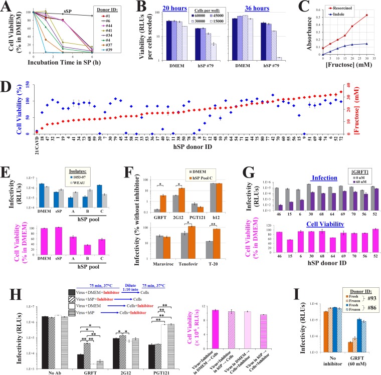
FIG 5.
HIV-1 infection and inhibition in human seminal plasma. (A) Dynamics of SP cytotoxicity. Cf2Th CD4+ CCR5+ cells were incubated with whole hSP from different donors or with sSP(+Fr) for the indicated times, washed, and cultured in DMEM for 36 h. Cell viability was measured by an ATP-based assay. (B) Cf2Th CD4+ CCR5+ cells were added to 96-well plates at the indicated densities. Samples were then incubated at 37°C for 75 min with DMEM or a sample containing 77.5% hSP and 22.5% Tris-saline buffer. All samples were then washed and further processed as described in Fig. 3C, and viability was measured after 20 or 36 h. Viability values are corrected for the number of cells seeded in each well. (C) Fructose concentrations measured by the resorcinol and indole assays. Fructose standards were diluted in Tris-buffered saline (pH 7.5). (D) Cell viability (after 75 min of exposure) and fructose content (quantified by resorcinol) in 59 hSP samples from different donors were measured. Since samples were tested in separate experiments, values are reported as a percentage of the viability in the DMEM control included in each assay. CAVD, congenital absence of the vas deferens. (E) Infection by virus containing strain 1053-07 or WEAU Envs in DMEM, sSP(+Fr), and three pools of donor hSP using the protocol shown in Fig. 3C. Cell viability was measured in the same experiment. (F) HIV-1 inhibition in hSP. Infection by virus that contains the WEAU Env was measured in DMEM and whole pooled hSP from 10 donors (pool C) in the presence of GRFT (60 nM), 2G12 (20 μg/ml), PGT121 (3 μg/ml), b12 (15 μg/ml), maraviroc (1 μM), tenofovir (2 μM), or T-20 (6 μM). Cell viability after exposure to hSP pool C was measured in the same experiment and was 59.8% of that measured in DMEM. Error bars, SEM. P values were determined by a two-tailed t test. *, P < 0.05; **, P < 0.005. (G) GRFT inhibition of virus containing the 1053-07 Env in hSP samples from different donors. Viability values are expressed as a percentage of those measured in samples incubated with DMEM. (H) (Left) Immobilized viruses containing the WEAU Env were treated as shown, using GRFT (50 nM), 2G12 (20 μg/ml), or PGT121 (3 μg/ml). Entry was then halted using trypsin and T-20, and infectivity was measured 3 days later. (Right) The viability of samples similarly treated in the same experiment was measured after 36 h. (I) Infection and GRFT inhibition in fresh and thawed hSPs. Samples were collected from donors and either subjected to one freeze-thaw cycle (frozen) or left at room temperature (fresh) for 4 h, before they were used for infection assays in the absence or presence of GRFT.

