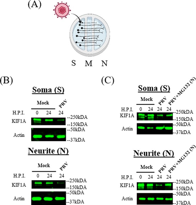
FIG 5.

PRV infection of compartmented primary neuronal cultures leads to reduction of KIF1A protein and proteasomal degradation in axons. (A) Illustration of in vitro culture of primary superior cervical ganglion neurons using modified Campenot chambers that include the S (soma), M (methocel), and N (neurite, axon termini) compartments. A total of 106 PFU of PRV Becker were added in the S compartment. (B and C) SCG neurons were infected at the S compartment with PRV Becker for 24 h. (B) Cell bodies and axons from the S and N compartments were collected at the time points shown, and lysates were analyzed by Western blotting to monitor the levels of KIF1A and actin. (C) DMSO or MG132 (2.5 μM) was added to the N compartment at 8 hpi. Cell bodies and axons from the S and N compartments were collected at the time points shown, and lysates were analyzed by Western blotting to monitor the levels of KIF1A and actin.
