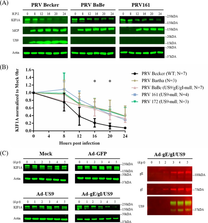
FIG 7.
PRV anterograde-spread complex US9/gE/gI promotes the accerlated degradation of KIF1A proteins. (A) Differentiated PC12 cells were infected with PRV Becker or different PRV mutants defective in anterograde spread (results from PRV BaBe and PRV161 infections are shown) for 0, 8, 12, 16, 20, or 24 h. Cells were harvested and lysates were analyzed by Western blotting to monitor KIF1A, MCP, US9, and actin protein levels. (B) For every infection, KIF1A levels were measured by band intensities and normalized with respect to actin levels at each time point. Normalized values were then normalized to those for mock-infected 0-h samples. Values are means plus SEMs (error bars) from seven independent experiments. *, P < 0.05 for all mutants versus PRV Becker at the specified time point. (C) Differentiated PC12 cells were transduced with adenoviral vectors expressing GFP, GFP-US9, or GFP-US9/gE-mCherry/gI-mTurquoise 2. Cells were collected at the indicated days posttransduction (d.p.t) and subjected to Western blot analysis to monitor KIF1A, gE, gI, US9, and actin levels.

