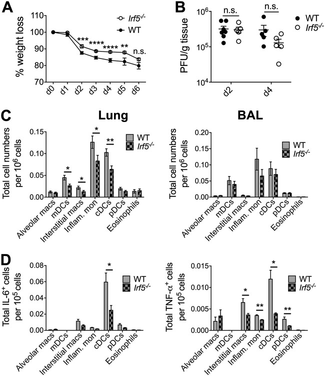
FIG 2.
IRF5 enhances influenza A virus-induced inflammatory response in a murine infection model. (A) Weight loss of WT and Irf5−/− mice was assessed over time, and comparable results were observed in 4 independent experiments, with 4 to 5 WT or Irf5−/− mice in each group per experiment. Data shown are the mean ± SEM. (B) Replication of virus in the lungs was quantified using a plaque assay. Data shown are the mean ± SEM using 7 WT and 5 Irf5−/− mice for day 2 and 5 mice of each genotype for day 4. (C) Recruitment of specific myeloid cell populations (mDCs, monocyte-derived DCs; cDCs, conventional DCs; pDCs, plasmacytoid DCs; Inflam. mon, inflammatory monocytes) in WT and Irf5−/− mice was assessed by flow cytometry 2 days p.i. Populations were defined by the following markers: alveolar macrophages (Alveolar macs), SiglecF+ CD11b+ CD64+ Ly6C−; mDCs, SiglecF− CD11b+ MHC-II+ CD11c+ CD64+ Ly6C+; interstitial macrophages, SiglecF− CD11b+ MHC-II+ CD11c− CD64+ Ly6C+; inflammatory monocytes, SiglecF− CD11b+ MHC-II− Ly6C+ CD64+; cDCs, MHC-II+ CD11c+ Ly6C−; pDCs, B220+ SiglecH+ MHC-IIlow CD11clow; and eosinophils, SiglecF+ CD11c− CD11b+ Ly6C−. Data shown are the mean ± SEM using 11 WT and 10 Irf5−/− mice from multiple replicates. (D) The total number of each individual myeloid cell population (unstimulated, ex vivo) positive for IL-6 and TNF-α expression was detected by flow cytometry, with data presented representing the mean total cell number per 105 cells of each cell type ± SEM. Data represent two experiments.

