
Figure 5.
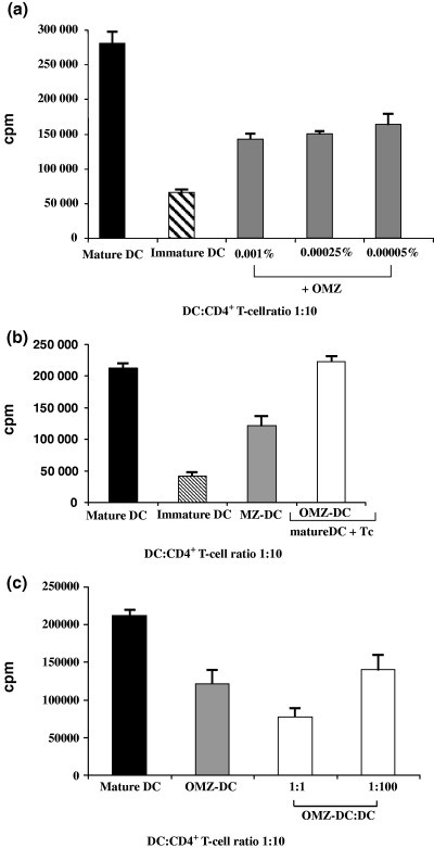
Reduced stimulatory capacity of oxymetazoline‐treated dendritic cells. (a) Freshly isolated CD4+ T lymphocytes (1 × 10⁵) were stimulated with allogeneic OMZ‐treated (0.001%) human DC (1 × 10⁴) in 96‐well plates for 4 days plus 16 h in the presence of 3[H]‐thymidine (37 kBq/well). Untreated mature and immature DC served as controls. The results of four independent experiments are shown. (b) CD4+ T cells (1 × 10⁵) were stimulated with allogeneic untreated DC (1 × 10⁴) in 24‐well plates. OMZ‐treated (0.001%) DC (1 × 10⁴) were placed in transwell chambers in the same well. After 4 days of culture, 3 × 200 μl per well of each culture were transferred to three individual wells of 96‐well plates. Proliferation was measured after an additional 16 h pulse with 3[H]‐thymidine. CD4+ T cells stimulated with untreated mature and immature DC and OMZ‐DC (0.001%) served as control. The results of three independent experiments are shown. (c) CD4+ T cells were stimulated with allogeneic untreated DC at a DC:T‐cell ratio of 1:10 and OMZ‐treated (0.001%) DC were added in increasing concentrations. Results of three independent experiments are shown.